
Overview of Bone Fractures

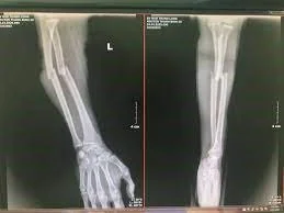
Lower leg fracture radiograph (X-ray) Forearm fracture radiograph (X-ray)
CAUSES.
– High-energy injuries: Traffic accidents, occupational accidents, and sports injuries,…
– Pathological fractures in the elderly: When bone quality deteriorates due to diseases such as osteoporosis, bone tumors, etc., or due to age-related bone density loss, the bones become more susceptible to stress and impact, easily leading to fractures even from very minor incidents such as improper posture or simple falls.
- –> leading to 2 types of injury: Bone fracture and soft tissue injury
- –> risk of compartment syndrome, delayed union, nonunion, postoperative infection, or even segmental bone loss, amputation, and loss of limb function.
CLASSIFICATION
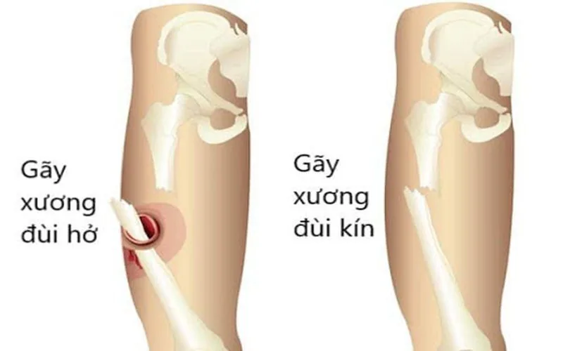
– Closed fracture and open fracture.
– Transverse fracture, oblique fracture, displaced fracture, comminuted fracture.
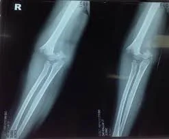
Transverse fracture Displaced fracture
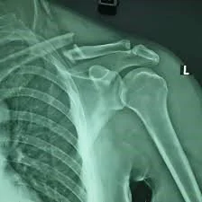
Displaced clavicle fracture with comminuted fragments.
CLINICAL SYMPTOMS.
– After the injury, the patient experiences some symptoms.
- Pain may be mild in hairline (fissure) fractures or sharp and intense in the affected limb, particularly during movement or when gripping objects.
- Limb deformity in cases of displaced fractures.
- Bruising, swelling, and redness at the site of bone injury.
- Possible presence of an open wound adjacent to the fracture site, accompanied by bleeding with visible fat droplets.
- Abnormal mobility and sounds (crepitus) at the fracture site.
- Restricted mobility or complete loss of limb function in the fractured area.
+ May experience shock, fainting due to pain, or blood loss.
WHAT TO DO WHEN A FRACTURE IS SUSPECTED?
- When signs of a suspected fracture appear, the patient should seek immediate assistance; absolutely avoid moving independently, as this may cause more severe trauma:
Dizziness due to pain, vascular and nerve damage caused by the displaced fracture end piercing.
- When witnessing someone suspected of having a bone fracture, we should:
- Reassure and calm the patient.
- Call for emergency medical assistance; during this time, temporarily immobilize the fractured limb, ideally with a sufficiently long wooden splint (if a wooden splint is unavailable, bamboo slats or any rigid materials may be used)...
rigid, load-bearing object may be adapted for temporary stabilization). Optimal immobilization of the fracture site significantly reduces the risk of secondary complications and facilitates
call for medical intervention afterwards.
- Loosen clothing, rings, bracelets, etc., around the affected limb to prevent constriction that could compromise blood supply to the extremity.
- If mild analgesics such as paracetamol are available, they may be administered to provide temporary pain relief while awaiting emergency medical support.
- Strictly avoid moving the patient before the fractured limb is adequately immobilized, do not extract foreign objects penetrating the fracture site, and do not wash or irrigate the wound...
open wounds directly, as this may induce neurogenic shock or provoke hemorrhage. Simply cover open wounds with a clean, damp cloth or sterile dressing.
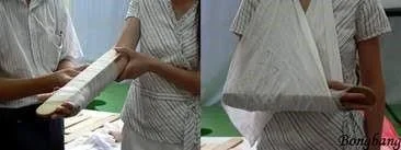
Wooden splint immobilization for fractured limb
HOW ARE FRACTURES DIAGNOSED?
– When you have clear signs or suspect a fracture, the doctor will examine you to further determine the risks of complications caused by the fracture and other dangerous injuries that may affect your life. Then, some tests will be conducted to diagnose:
- X-ray: Visualizes the location, fracture pattern, and degree of displacement.
- CT scan: Provides a more comprehensive evaluation of the fracture site, especially for intra-articular and peri-articular fractures, to establish an optimal treatment plan.
- MRI: Commonly utilized in diagnosing hairline or stress fractures occult on X-rays, or suspected pathological fractures associated with bone tumors, bone tuberculosis, osteomyelitis, etc.
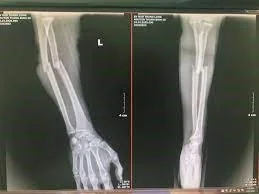
Forearm fracture X-ray
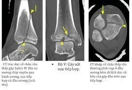
CT scan of distal tibial fracture

Stress fracture
HOW TO TREAT FRACTURES?
– Depending on the fracture shape on the X-ray and the extent of soft tissue damage in the fractured limb, the doctor will develop a detailed treatment plan. Therefore, diagnosis and treatment must be performed by an experienced specialist doctor.
– Current treatment methods:
- Casting: Utilizes plaster of Paris or fiberglass casting to immobilize the fracture site for several weeks. The primary advantage is non-surgical management; however, it carries considerable risks of complications such as compartment syndrome, delayed union, muscle atrophy, joint stiffness, and pseudoarthrosis (nonunion). Therefore, casting is performed strictly when indicated or when the patient is not a candidate for surgical osteosynthesis (bone fixation).
- Orthopedic braces and splints: Figure-of-eight brace for clavicle fractures, Desault bandage for proximal humerus fractures, Iselin splint for fractures of metacarpals, metatarsals, fingers, and toes, etc.
- External frame fixation
- Internal fixation (Osteosynthesis): Plates and screws, intramedullary nails, Kirschner wires (K-wires), cancellous screws, etc.
– Orthopedic surgery will help patients return to work and care for themselves sooner, relieving the affected joints and thereby reducing the risk of muscle atrophy and joint stiffness. However, there are still risks associated with the intervention, such as bone membrane grafting to place bone fixation devices. Therefore, patients should visit specialized medical facilities with experienced specialists and adequate resources to enhance treatment quality.
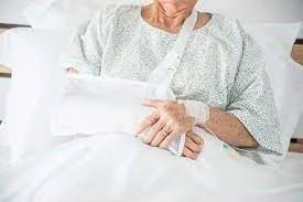
Đeo đai Sling Bó bột
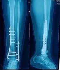
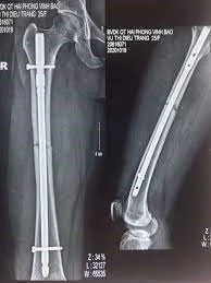
Plate and screw fixation Intramedullary nailing
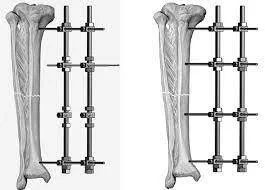
External frame fixation
STRATEGIES FOR BONE FRACTURE PREVENTION
– A reasonable supplementary nutrition regimen: To enhance bone strength, it is necessary to ensure the body receives 1200 – 1500 milligrams (mg) of calcium and 800 - 1000 international units (IU) of vitamin D daily. Calcium and vitamin D can be supplemented by:
- Enhance your daily nutrition with calcium-rich foods to maintain robust bone health, especially for postmenopausal women and individuals over 35 years of age. Calcium is abundantly found in foods such as milk, yogurt, dairy products, and dark green leafy vegetables…
- Boost vitamin D intake to optimize calcium absorption. Vitamin D can be obtained through natural sunlight exposure, oily fish, eggs, etc.
– Daily activities.
- Healthy lifestyle habits and physical activities also play a vital role in preventing fractures:
. Perform weight-bearing exercises to improve muscle mass and bone density, such as walking, jogging, weight training, swimming...
. Perform physical exercises and physiotherapy to enhance balance.
. Reduce the risk of falls: Keep your home tidy, use non-slip mats, watch your step, use walking aids (if necessary)...
. Be cautious during work, daily activities, and traffic participation to reduce the risk of injuries.
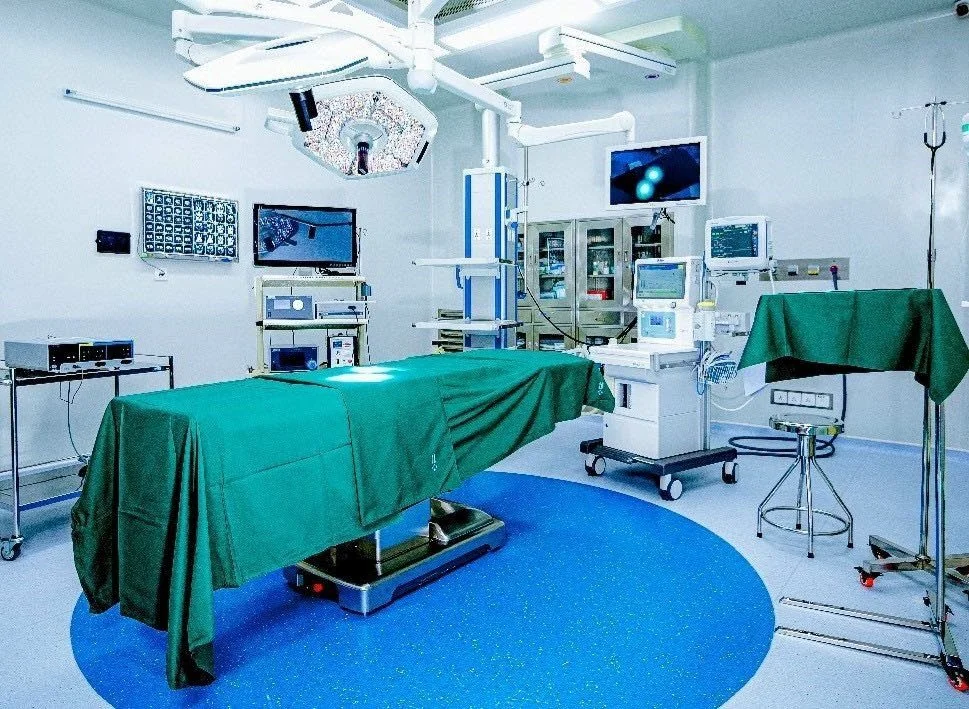
Proactively protect your health with our experienced specialist doctors and advanced medical equipment.
📞 BOOK AN APPOINTMENT NOW: (+84) 28 2200 8686 – (+84) 901 295 399
⏰ Monday – Saturday: Morning 7:30 AM – 12:00 PM, Afternoon 1:30 PM – 5:00 PM | 24/7 Emergency: ((+84) 937 389 539)
💳 Full nationwide health insurance coverage & Direct billing for private insurance accepted